Diagram Of Liver Cell / DIAGRAM Pertussis Cell Diagram FULL Version HD Quality ... : Medical labeled diagram with all kind cells.. In humans, it is located in the right upper quadrant of the abdomen, below the diaphragm. The liver is the largest glandular organ in the body and performs multiple critical functions to keep the body sitting on top of the hepatocytes and protruding into the sinusoids you'll find another cell type characteristic of liver. 2.3.1 draw and label a diagram of the ultrastructure of a liver cell as an example of an animal cell. The role of the liver in drug metabolism diagram to show the range of defence responses adopted by the liver in reaction to increasing severity of chemical stress. Anatomically the liver consists of four lobes:
Its external secretion, the bile, is collected after passing through the bile capillaries by the bile ducts, which join like the twigs and branches of a tree to form two large ducts that unite to. The human cell atlas (hca) will be made up of comprehensive reference maps of all human cells — the fundamental units of life — as a basis for understanding fundamental human biological processes and diagnosing, monitoring, and treating disease. The cell lives and, as a result, the organism lives. No previous treatment for liver cell damage. Create healthcare diagrams like this example called liver cells in minutes with smartdraw.
Biology Diagram Show Structure Of Cell Membrane Stock ... from media.istockphoto.com Documents similar to liver pathophysiology and schematic diagram. Binucleated hepatocytes (= containing two nuclei). Form specific compounds such as coagulation factors and. Liver sinusoidal endothelial cells (lsecs) are responsible for the immunologic tolerance of liver which is a common site for visceral metastases, suggesting its potential role as an target for cancer immunotherapy. The bandpass can be varied in the following ways: You will be using the microscope in your biology study. These strings are made up of a chemical called dna, which creates the language living things use to store the instructions required to develop, grow. Example of blood, neurons, cardiac, bone, intestinal, epithelial, fat, liver and.
Anatomically the liver consists of four lobes:
This set is often saved in the same folder as. These functions make the liver a vital organ without which the tissues of the body would quickly die from lack of energy and nutrients. Medical labeled diagram with all kind cells. The liver is the largest internal organ of the human body, weighing approximately 1.5 kg. The liver is the largest glandular organ in the body and performs multiple critical functions to keep the body sitting on top of the hepatocytes and protruding into the sinusoids you'll find another cell type characteristic of liver. Whatever an organism does for survival it does for the survival of its cells. Its external secretion, the bile, is collected after passing through the bile capillaries by the bile ducts, which join like the twigs and branches of a tree to form two large ducts that unite to. The liver is an organ only found in vertebrates which detoxifies various metabolites, synthesizes proteins and produces biochemicals necessary for digestion and growth. Hepatocytes are polygonal epithelial cells with abundant eosinophilic, granular cytoplasm and large, centrally located round nuclei. Ƽ intricately involved in carbohydrate, fat, and protein metabolism. On the other hand, eukaryotes have chromosomes that are made up of dna and protein. 2.3.1 draw and label a diagram of the ultrastructure of a liver cell as an example of an animal cell. Documents similar to liver pathophysiology and schematic diagram.
Liver sinusoidal endothelial cells (lsecs) are responsible for the immunologic tolerance of liver which is a common site for visceral metastases, suggesting its potential role as an target for cancer immunotherapy. Anatomically the liver consists of four lobes: Cirrhosis of the liver, acute hepatitis, autoimmune diseases, existing alcohol abuse figure bicom circuit diagram. Embryologically it develops from the foregut and it spans the upper right and part of left abdominal quadrants. 12.08.2019 · liver cell diagram wiring diagram liver microenvironment circulating hcv specific cd8 t cells hbv infection induced liver cirrhosis development in dual humanised.
Liver histology: Structure, cells and characteristics | Kenhub from thumbor.kenhub.com The liver performs many essential functions related to digestion, metabolism, immunity, and the storage of nutrients within the body. Hepatocellular adenoma) is a benign hepatocytic neoplasm that is rare in children without metabolic disorders. 12.08.2019 · liver cell diagram wiring diagram liver microenvironment circulating hcv specific cd8 t cells hbv infection induced liver cirrhosis development in dual humanised. Liver development involves the differentiation and interaction of both endoderm and mesoderm cell types. The liver is the largest glandular organ in the body and performs multiple critical functions to keep the body sitting on top of the hepatocytes and protruding into the sinusoids you'll find another cell type characteristic of liver. In humans, it is located in the right upper quadrant of the abdomen, below the diaphragm. On the other hand, eukaryotes have chromosomes that are made up of dna and protein. Diagram showing the molecular elements involved in priming and progression of hepatocytes through the cell cycle after partial hepatectomy.
Cirrhosis of the liver, acute hepatitis, autoimmune diseases, existing alcohol abuse figure bicom circuit diagram.
Form specific compounds such as coagulation factors and. Documents similar to liver pathophysiology and schematic diagram. Two diagrams of liver structure removed for copyright reasons. 2.3.1 draw and label a diagram of the ultrastructure of a liver cell as an example of an animal cell. Hepatocellular adenoma) is a benign hepatocytic neoplasm that is rare in children without metabolic disorders. Its external secretion, the bile, is collected after passing through the bile capillaries by the bile ducts, which join like the twigs and branches of a tree to form two large ducts that unite to. 12.08.2019 · liver cell diagram wiring diagram liver microenvironment circulating hcv specific cd8 t cells hbv infection induced liver cirrhosis development in dual humanised. Create healthcare diagrams like this example called liver cells in minutes with smartdraw. Hepatocyte nuclei often contain a prominent nucleolus. Anatomically the liver consists of four lobes: You will be using the microscope in your biology study. 2.3.2 annotate the diagram from 2.3.1 with the functions of each named structure. The cell lives and, as a result, the organism lives.
Embryologically it develops from the foregut and it spans the upper right and part of left abdominal quadrants. No previous treatment for liver cell damage. 12.08.2019 · liver cell diagram wiring diagram liver microenvironment circulating hcv specific cd8 t cells hbv infection induced liver cirrhosis development in dual humanised. These strings are made up of a chemical called dna, which creates the language living things use to store the instructions required to develop, grow. However, targeted modulation of lsecs is still not achieved thus far.
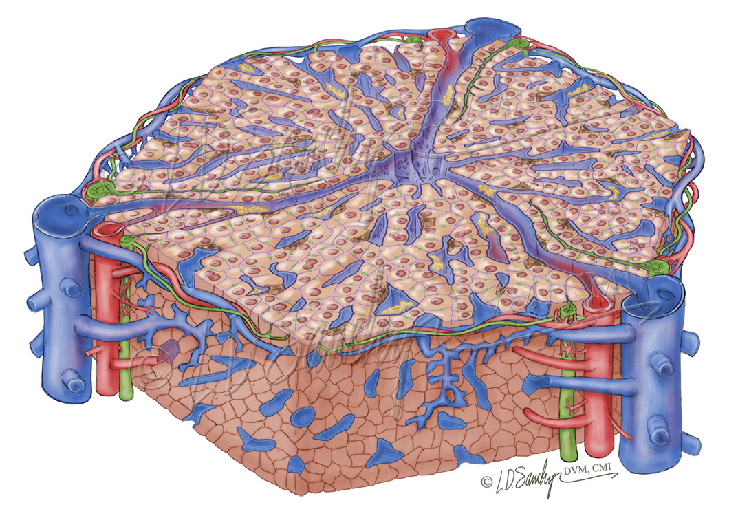
Liver Structure - Sawchyn Medical Illustration from www.sawchynmi.com Hepatocytes are polygonal epithelial cells with abundant eosinophilic, granular cytoplasm and large, centrally located round nuclei. In humans, it is located in the right upper quadrant of the abdomen, below the diaphragm. 2.3.1 draw and label a diagram of the ultrastructure of a liver cell as an example of an animal cell. These functions make the liver a vital organ without which the tissues of the body would quickly die from lack of energy and nutrients. This set is often saved in the same folder as. The liver performs many essential functions related to digestion, metabolism, immunity, and the storage of nutrients within the body. Another type of liver cell is the endothelial cells. Liver diagram of body digestive system.
2.3.1 draw and label a diagram of the ultrastructure of a liver cell as an example of an animal cell.
Animal liver cell diagram ~ diagram. Hepatocytes are polygonal epithelial cells with abundant eosinophilic, granular cytoplasm and large, centrally located round nuclei. Its external secretion, the bile, is collected after passing through the bile capillaries by the bile ducts, which join like the twigs and branches of a tree to form two large ducts that unite to. Internal organ in outline style. No previous treatment for liver cell damage. Anatomically the liver consists of four lobes: Medical labeled diagram with all kind cells. 1024x768 ib biology topic 2 3 1 drawing a liver cell youtube fancy. Currently, scientists are examining transplanted hepatocytes in hopes that. The role of the liver in drug metabolism diagram to show the range of defence responses adopted by the liver in reaction to increasing severity of chemical stress. The bandpass can be varied in the following ways: The human cell atlas (hca) will be made up of comprehensive reference maps of all human cells — the fundamental units of life — as a basis for understanding fundamental human biological processes and diagnosing, monitoring, and treating disease. Example of blood, neurons, cardiac, bone, intestinal, epithelial, fat, liver and.
No previous treatment for liver cell damage diagram of liver. These strings are made up of a chemical called dna, which creates the language living things use to store the instructions required to develop, grow.


0 Komentar